MAGNETIC RESONANCE IMAGING (MRI)
MRI is an imaging technique which provides very detailed pictures of the internal structure of the various parts of the body, including the brain. The machine uses a strong magnet and radiofrequency magnetic field (a magnetic field with a frequency similar to your FM radio) to make images of the body interior.
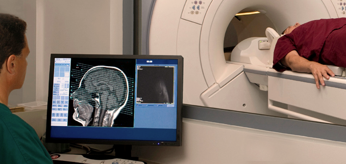
To be scanned, you will first lie down on a long narrow bed which is then moved into the center of the scanner so that the machine can gather data. During this time you will be exposed to a radiofrequency magnetic field; you will NOT be exposed to any x-rays. You will, however, hear loud repetitive tapping and grinding noises which arise from the magnetic coils in the machine. You will be provided with earplugs to muffle the sound.
In order to image the brain clearly it is necessary for the head to be held very still while lying in the scanner. To make this easier we may place foam wedges around the head, use Velcro straps, or some special tape to ensure that there is no movement.
The magnetic fields are not known to cause harmful effects at the levels used in the scanner. Federal guidelines have been developed for these machines; these guidelines will be followed. MR scanning will be conducted by qualified personnel. As metallic objects may experience a strong attraction to the magnet, it is very important that you notify the researcher of any metal objects, devices, or implants that are in or on your body before entering the magnet room. This includes metal for medical purposes as braces or pacemakers, prostheses, and other metallic objects imbedded in the body such as bullets, buckshot, shrapnel, and any metal fragments from working around metal. All other metallic objects must also be removed prior to entering the magnet room including keys, jewelry, pocket knives, money clips, paper clips, safety pins, hair pins, and barrettes. In addition, objects such as watches, credit cards, and hearing aids could be damaged in the presence of the magnetic field.
SUPPORT